Lameness is an abnormality of the horse’s gait that could be caused by pain, a mechanical problem such as “stringhalt” (muscle spasms), or a neurological problem such as “wobblers”.
Some causes of equine lameness:
- Abscesses in the foot
- Hoof Wall Cracks
- Inflammation of the foot
- Strain in tendons or joints
- Bone chips
- Fractures
- Arthritis (inflamed joints)
- Back pain
- Nerve damage
- Muscle soreness
- Wounds, cuts, and bruises
Visual Examination
Examine the animal visually at a distance; note the body type and condition, conformation, any shifting in weight or abnormal stances, and the attitude of the animal.
Have a closer visual examination of the animal. Look for abnormal wear in the feet, cracks in hoof, lacerations, swellings in joints or tendons, atrophy or swelling of the muscles, and any other gross changes.
An Animal can also be observed during exercise, at the walk, trot, and sometimes at the canter or gallop. Mostly the trot is the most helpful gait for the exam because of its symmetry.
The examination should be done by observing the horse movement from the front, the side and from behind. Also, circling the horse or moving the horse in figure eight (8) can locate lameness.
Observe head nodding, gait deficits, alterations in the height of the foot flight arc, phase of stride, joint flexion angle, foot placement, and symmetry in gluteal rise and duration.
Palpation and Manipulation
Observe the size and shape of the foot. Compare the normal with the abnormal. Look for any abnormal hoof wear, ring formation, heel bulb contraction, hoof wall cracks and swellings, and any other asymmetries. Palpate the coronary band for heat, swelling and pain on pressure.
Lameness due to Hoof Wall Damage
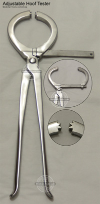
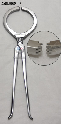
Hoof Wall Cracks may be associated with lameness. This could be checked with hoof tester examination. The examination applies a hoof tester in a systematic manner, to the entire sole and frog region and hoof wall. When applied properly, this instrument allows the examiner to palpate the hoof. Medical Tools Adjustable Hoof Tester 16” new design allows adjustable jaws over entire hoof surface; longer handle provides extra leverage. Jaws remain parallel for more even and steadier diagnostic pressure,
For a sensitive horse, the examiner may need to use gentle application at first, followed by firmer pressure. The examiner is trying to identify and localize hoof sensitivity. Although this sounds simple, it requires experience.
The arm of the hoof tester that is applied to the hoof wall needs to be continually checked so pressure is not being applied to the coronet hand. I prefer to begin at the lateral or medial angle of the sole and continue examination by applying hoof tester pressure every 2 to 3cm until the entire surface of the sole has been checked.
Next apply pressure to the frog (caudal, central and carnial) then I use the hoof tester on the hoof wall at the heels. Finally, I apply the tester diagonally from the medial heel to the dorsolateral hoof and from the lateral heel to the dorsolateral hoof. The diagonal application is probably most useful for the horses with pads.
If sensitive is encountered, it is absolutely necessary to confirm that the examiner has revealed true sensitivity by the horse. True sensitivity is identified by repeated intermittent hoof tester pressure that results in persistent reflexive withdrawal (flexing the shoulder) obviously, different amount of hoof tester pressure are applied to elicit a response, depending on the sole thickness and the painfulness of the condition. Hoof Tester response should be compared with those obtained from opposite foot.
Hoof Testers


